Benefits of Cone Beam CT Scans
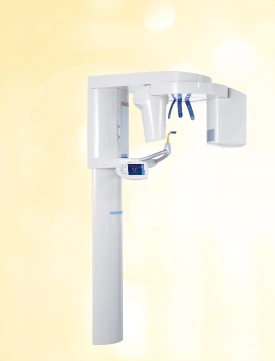
Welcome to Green Dental, Sherman Oaks, your trusted dental care provider committed to delivering exceptional services using the latest technology. We are proud to offer i-CAT® 3D Imaging, a revolutionary dental imaging system that allows for precise and comprehensive diagnostics.
We understand the importance of accurate & detailed imaging in dentistry. With i-CAT® 3D Imaging, our dental team can capture high-resolution 3D images of your teeth, jaw, and surrounding structures. This advanced technology enables our highly skilled dentists to have a comprehensive view of your oral health, aiding in accurate diagnoses and effective treatment planning.
In our dental practice, we use the most advanced 3D CT technology, which provides highly accurate 3-D radiographs that allow us to diagnose your oral surgery, plan a surgical course of treatment, and help you heal.
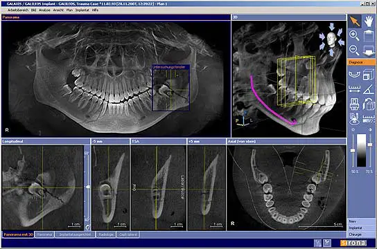
3-D CT scans provide highly accurate images of the mouth. These images allow dentists to diagnose potential problems such as TMJ, analyze an airway, and plan surgeries. They are also excellent for diagnosing and treating dental implantology, orthognathic surgery, and other dental problems.
With the help of 3-D CT scans, undistorted, anatomically correct views of the jaws, teeth and facial bones along with cross-sectional (bucco-lingual), axial, coronal, sagittal, cephalometric and panoramic views are generated quickly and painlessly.
3D imaging enables a level of anatomical accuracy, therapeutic ability and patient care not possible with 2D technologies.
3D imaging can be used pre-surgically to plan the surgical site and to plan the appropriate incision technique, it is also useful in neurological, orthopedic, reproductive, ophthalmic and otolaryngologic surgery as well as endovascular intervention surgeries.
With the addition of cone-beam CT technology in our office, our dental practice is committed to providing innovative, high-quality, patient care.
i-CAT® 3D Imaging
- Dental implants
- Location of anatomic structures: mandibular canal, submandibular fossa, incisive canal, maxillary sinus
- Size and shape of ridge, quantity and quality of bone
- Number, orientation of implants
- Need for bone graft, sinus lift
- 3D CT ScanUse of implant planning software
- Oral and maxillofacial surgery
- Relationship of third molar roots to mandibular canal to minimize any nerve damage
- Localization of impacted teeth, foreign objects, extra teeth
- Evaluation of facial fractures and asymmetry
- Orthognathic surgery planning
- Oral and maxillofacial pathology
- Localization and characterization of lesions in the jaws
- Effect of lesion on jaw in 3rd dimension: expansion, cortical erosion, bilateral symmetry
- Relationship of lesion to teeth and other structures